Anatomy of the muscles of the neck and head of a person: structure and function. Middle group of muscles of the neck Superficial muscles of the neck
The platysma muscle of the neck (platysma) tightens the skin of the neck and part of the sternum, and also shifts the angle of the mouth forward and downward. The muscle is a thin, wide plate located under the skin of the neck and partly under the skin of the face. The point of its beginning is in the subclavian region of the fascia of the pectoralis major and deltoid muscles, and the edge of the lower jaw, the chewing fascia and the corner of the mouth serve as the attachment point.
The sternocleidomastoid muscle (m. Sternocleidomastoideus) (Fig. 90, 95, 96), with a bilateral contraction, throws its head back, and with a one-sided contraction, it tilts its head to its side (in the direction on which the muscle contracts) and turns it in the opposite direction.
The muscle is a thick long cord with two heads, going obliquely from the mastoid process through the neck to the sternoclavicular joint. The lateral head of the muscle has the starting point of the anterior surface of the handle of the sternum, and the medial head has the sternal end of the clavicle. The muscle is attached to the mastoid process and the lateral part of the superior nuchal line.
 | Rice. 96. Superficial, median and deep muscles of the neck (side view): 1 - stylohyoid muscle; |
 | Rice. 97. Median and deep muscles of the neck (side view): 1 - maxillary-hyoid muscle; |
The muscles of the neck have a very specific structure, which is associated with their origin, functionality, relationships with the circulatory system, blood vessels, nerve endings and internal organs.
Human anatomy has subdivided this tissue into several important groups: superficial, median, and deep. The middle group includes muscle tissues that are located above and below the hyoid bone. Deep muscle tissue is usually divided into lateral and medial groups.
News line ✆
Human anatomy classifies the superficial muscles of the neck into several types:
- subcutaneous muscle of the neck, which looks like a thin wide plate under the skin and some part of the face. The subcutaneous muscle of the neck begins in the subclavian sector from the fascia of the pectoralis major, deltoid, and is attached to the corner of the mouth, the edge of the lower jaw. Performs the functions of lifting the skin, skin in the chest area, pulls the corners of the mouth downward and outward;
- The sternocleidomastoid is a long and thick strand that crosses the neck obliquely, from the mastoid process to the sternoclavicular joint. These superficial neck muscles have two heads. They serve to turn the head to the opposite side, tilt it;
- the muscle of the neck has an oblong shape and begins near the processes of the III-VII cervical vertebrae, the bodies of the first thoracic vertebrae, is attached to the lateral sectors of the upper line. She pulls the neck back, provides rotation of this spinal section in her direction.

Middle group
The median neck muscles include:
- the digastric muscle consists of two abdomens, which are connected by a tendon bridge. They start from the fossa of the lower jaw, as well as a specific cut of the temporal sector, and end with an attachment to the tendon, which is attached to the hyoid body of the bone. The main function is to lower and pull the lower part of the jaw back;
- The stylohyoid is a fusiform muscle tissue. It originates from the process of the styloid form of the temporal bone, attaches to the body of the hyoid bone. She pulls the hyoid bone upward, backward and outward;
- the jaw-hyoid connects with the same muscle tissue on the opposite side and forms the floor of the oral cavity. They are located on the line of the lower jaw, fixed to the front side of the same bone and form the maxillary-hyoid suture of the diaphragm of the mouth;
- the sublingual is located above the sublingual, attaches to the anterior sector of the body of the hyoid bone;
- The scapular-hyoid intermediate tendon ligament divides into two abdomens. It is located in the lower edge of the hyoid body of the bone, the upper edge of the scapula. The main function is to pull the hyoid bone downward and outward while fixing the scapula;
- The sternohyoid originates in the posterior sector of the clavicle, the handle of the sternum, is attached to the lower edge of the body of the hyoid bone;
- The sterno-thyroid originates from the posterior region of the sternum, is fixed by the oblique line of the thyroid cartilaginous tissue of the larynx;
- the thyroid-hyoid serves to bring the hypoglossal body closer to the bone and larynx while they are in a state of fixation.

Deep group
Deep neck muscles are divided into different types of scalene:
- the anterior scalene is located near the anterior tubercles of the 3-6 cervical vertebrae, attached to the tubercle of the anterior scalene 1 rib. Plays an important role in the process of contraction of the cervical spine in its direction, tilts it forward;
- the middle scalene begins near the anterior tubercles of the lower cervical vertebral bodies, is fixed in the upper region of 1 rib. The main role is to raise the rib, as well as tilt the cervical region forward;
- posterior scalene is formed in the posterior tubercles of 4-6 cervical vertebrae, attached to the outer region of 2 ribs. Functional feature - raising the rib, bending the cervical spine forward while fixing the chest;
- the long neck muscle includes two parts - the lower and the upper. Their base starts from the bodies of the first three vertebrae of the thoracic region, the processes of 4-6 bodies of the cervical vertebrae. Attachment occurs in bodies 2-4 and transverse processes and the anterior tubercle of the 1st vertebra. The long neck muscle serves to tilt it forward to its side;
- the long muscle of the head goes from the anterior tubercles of the III-VI cervical vertebrae and is attached to the lower surface of the main bone of the occiput;
- the lateral straight line originates at the base of the 1st cervical vertebra, is attached to the lateral part of the occiput bone, while performing the function of tilting the head.

The neck muscles play an important role in the formation of the muscular corset of both individual vertebrae and the spine as a whole.
Muscles serve as a kind of armor that protects the internal organs of the head, bone tissue, and also forms the upper sections of the muscular corset for the spine and the whole body.
It is extremely important to monitor the state of this part of the human body in order to avoid various pathologies that can affect the work of the whole organism. Various prophylaxis, timely diagnostics, attentive and careful attitude to your body are the key to long and good health.
No need to treat joints with pills!

Have you ever experienced unpleasant joint discomfort, annoying back pain? Judging by the fact that you are reading this article, you or your loved ones are faced with this problem. And you know firsthand what it is.
The superficial muscles of the neck lie in two layers.
1. Subcutaneous muscle of the neck ( platysma), is a wide thin quadrangular plate; starts from the skin of the upper chest at the level of 2-3 ribs, rises up along the neck; its medial fibers attach to the lower jaw, lateral fibers continue into the muscles of the mouth. Function: tightens the skin of the neck, lowers the corner of the mouth, promotes the outflow of blood from the superficial veins of the neck.
2. Sternocleidomastoid muscle
(m. sternocleidomastoideus), located under the subcutaneous muscle of the neck, begins with two heads. The sternal head originates from the upper part of the anterior surface of the sternum, the clavicular head - from the upper surface of the medial third of the clavicle, the muscle is attached to the lateral half of the superior nuchal line and the lateral surface of the mastoid process of the temporal bone. Function: with one-sided contraction, tilts the head to the shoulder, while the face turns in the opposite direction; with a bilateral contraction, it holds the head in an upright position; with a strong contraction, it throws back the head, since the place of muscle attachment is located behind the frontal axis of the atlanto-occipital joint; with a fixed head, it can participate in lifting the girdle of the upper limb and chest (inspiratory accessory muscle).
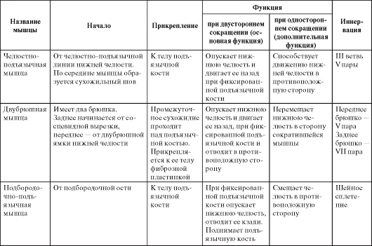
Middle neck muscles (muscles of the hyoid bone)
Suprahyoid muscles (musculi suprahyoidei)
1. Digastric muscle ( m. digastricus), has two abdomens - anterior and posterior. The anterior abdomen (venter anterior) starts from the fossa digastrica at the base of the lower jaw, goes back and down. The posterior abdomen (venter posterior) starts from the incisura mastoidea, goes forward and downward. Both abdomens are connected by an intermediate tendon, which pierces the stylohyoid muscle (m. Stylohyoideus) and, through a fibrous loop, is attached to the body and the great horn of the hyoid bone. Function: lowers the lower jaw, with a fixed lower jaw raises the hyoid bone, the posterior abdomen participates in swallowing and chewing.
2. Stylohyoid muscle (m. Stylohyoideus) , begins with a small tendon from the posterior surface of the styloid process near its base, goes forward and downward, attaches to the body of the hyoid bone next to the great horn just above the attachment pointscapular-hyoidmuscles (see below). Near
the insertion site of the muscle pierces the intermediate tendon of the digastric muscle. Function: lifts the hyoid bone and pulls it back.
3. Maxillofacial muscle (m. Mylohyoideus) , lies above the anterior abdomen of the digastric muscle. Starts from linea mylohyoidea of the lower jaw, the posterior fibers are directed slightly downward and medially and are attached to the body of the hyoid bone, the anterior and middle fibers are connected to the same fibers of the opposite side, forming a fibrous suture along the midline that stretches from the middle of the chin to the hyoid bone. Right and leftmaxillary-hyoidthe muscles, together with the anterior abdomens of the digastric muscles, form the floor of the oral cavity ( diaphragma oris). Functions : raises the floor of the mouth in the first phase of swallowing, can raise the hyoid bone and lower the lower jaw.
4. Chin-hypoglossal muscle (m. Geniohyoideus) , lies above the medial partmaxillary-hyoidmuscles, starts from spina mentalis , goes back and slightly down, attaches to the anterior surface of the body of the hyoid bone. The right and left sublingual muscles sometimes merge with each other and with the sublingual muscle (one of the muscles of the tongue lies above m. geniohyoideus, starts from spina mentalis , goes into the thickness of the tongue)... Function : raises the hyoid bone and pulls it forward, while acting as an antagonist of the stylohyoid muscle; when the hyoid bone is fixed, the lower jaw drops.
Subhyoid muscles (musculi infrahyoidei)
1. Sternohyoid muscle (m. Sternohyoideus) - a thin narrow muscle, starts from the posterior surface of the sternal end of the clavicle, the posterior surface of the sternum handle and from the posteriorsternoclavicularligament, rises up and is attached to the lower surface of the body of the hyoid bone. Below there is a gap between the right and left muscles, at the top they converge to the midline.Sternohyoidmuscle may be absent, may be double, may be supplementedclavicular-sublingualmuscle or interrupted by tendon bridges. Function : lowers the hyoid bone, if it was raised before.
2. Sterno-thyroid muscle (m. Sternothyroideus) , shorter and wider than the previous one, lies deeper and slightly more medial to it; starts from the back surface of the sternum handle below m. sternohyoideus and from the posterior surface of the cartilage of the first rib, attaches to the oblique line of the laryngeal thyroid cartilage plate. Function : pulls the larynx down
after being lifted by swallowing or vocal movements.
3. Shield-hypoglossal muscle (m. Thyrohyoideus) , a small quadrangular muscle, which can be considered an upward extension of the previous muscle. It starts from the oblique line of the thyroid cartilage, attaches to the lower edge of the body and the large horn of the hyoid bone. Function : lowers the hyoid bone; when the hyoid bone is stabilized, it lifts the larynx upward.
4. The scapular-hyoid muscle (m. Omohyoideus) , consists of two abdomens - upper and lower, connected at an angle by an intermediate tendon. Lower abdomen ( venter inferior ) has the shape of a narrow flat strip that starts from the upper edge of the scapula medially incisura scapulae and from the superior transverse ligament of the scapula, goes forward and upward behindsternocleidomastoidmuscles and passes into the intermediate tendon. Upper abdomen ( venter superior ), starts from the intermediate tendon, rises vertically upward, attaches to the lower edge of the body of the hyoid bone, lateralsternohyoidmuscles. The intermediate tendon loops around a bundle of fibers of the deep fascia of the neck, which attaches to the clavicle and the first rib, maintaining the angular shape of the muscle. Function : drops the hyoid bone after it has been lifted; stretches its own fascia of the neck, while expanding the lumen of the deep cervical veins, the wall of which is fused with the fascia.
Deep neck muscles
Lateral (scalene) muscles
The scalene muscles of the neck correspond to the intercostal muscles of the chest, they start from the transverse processes of the cervical vertebrae and attach to the first (anterior and middle) and second (posterior) ribs.
1. Anterior scalene muscle ( m. scalenus anterior), starts from
anterior tubercles of the transverse processes C3-6, attached to tuberculum m. sacleni anterioris the first rib.
2. The middle scalene muscle ( m. scalenus medius), starts from
transverse processes C1 (2) –7, attached to the first rib, behind the sulcus arteriae subclaviae.
3. The posterior scalene muscle ( m. scalenus posterior), starts from
posterior tubercles C5–7, attached to the second rib.
Function: scalene muscles have "floating" punctum fixum and
punctum mobile; if punctum mobile is located on the transverse processes
vertebrae, then the scalene muscles tilt the cervical spine to their side, if the punctum mobile is on the ribs, then the scalene muscles lift the first and second ribs, acting as inhalation muscles.
Prevertebral muscles
1. Long muscle of the head ( m. longus capitis) , lies most superficially, starts from the anterior tubercles of the transverse processes
C3–6, attaches to the pars basilaris of the occipital bone. Function: bends the cervical spine, tilts the head forward.
2. Long neck muscle ( m. longus colli) , lies deeper than the previous muscle, has the shape of a triangle with the base facing medially, lies on the front surface of the bodies of all cervical and three upper thoracic vertebrae, consists of three parts: vertical, upper oblique and lower oblique.
∙ The vertical part is located medially (forms the base of the triangle), starts from the front surface of the bodies of the three upper
thoracic (Th1–3) and three lower cervical (C5–7) vertebrae, attaches to the anterior surface of the bodies of the three upper cervical vertebrae (C1–3).
∙ The lower oblique part is located laterally and below, begins
on the anterior surface of Th1–3 bodies, attaches to the anterior tubercles
C 5-7.
∙ The upper oblique part is located laterally and from above, begins
from the anterior tubercles of the transverse processes C2-5, attaches to the anterior tubercle of the atlas and the body of the second cervical vertebra.
Function: bends the cervical spine with bilateral contraction
the spinal column, with one-sided - tilts it in its direction. When contracting the upper oblique part, it turns the neck to its side, while contracting the lower oblique part, it turns the neck in the opposite direction.
3. Anterior rectus muscle of the head (m. Rectus capitis anterior)
starts from the lateral mass of the atlas, attaches to the basilar part of the occipital bone. Function: tilts the head forward, acting on the atlanto-occipital joint.
4. Lateral rectus muscle of the head (m. Rectus capitis lateralis)
starts from the transverse process of the atlas, attaches to the jugular process of the occipital bone. Function: tilts the head to its side, acting on the atlanto-occipital joint.
1.M. platysma, subcutaneous muscle of the neck, lies directly under the skin on the fascia in the form of a thin plate. It starts at the level of the II rib from the fascia pectoralis et deltoidea and attaches to the edge of the lower jaw and to the fascia parotfdea et fascia masseterica, partly continuing into the muscles of the mouth. (Inn. N. Facialis.)
Function. Pulling the skin of the neck, the muscle protects the saphenous veins from compression; in addition, she can pull down the corner of the mouth, which is important in facial expressions.
2. M. sternocleidomastoides, sternocleidomastoid muscle, lies immediately under the previous one, separating from it by the cervical fascia. It starts from the handle of the sternum and from the sternal end of the clavicle and attaches to the mastoid process and to the linea nuchae superior of the occipital bone. By its origin, the muscle is the detached part of m. trapezius and therefore has one innervation with this muscle (n. accessorius and CII).
Function. With a unilateral contraction, the muscle tilts the cervical spine towards its side; at the same time, the head is raised with a rotation of the face in the opposite direction.
With bilateral contraction, the muscles hold the head in an upright position, therefore the muscle itself and the place of its attachment (processus mastoideus) are most developed in humans in connection with upright posture. With bilateral contraction, flexion of the cervical spine can also occur with simultaneous lifting of the face. When fixing the head, it is possible to lift the chest during breathing (inspiratory accessory muscle).


With fractures of the lower jaw, the function of each of the masticatory muscles is realized differently than normal, and depends on how the fracture line passes. So, if the fracture line passes through the neck of the lower jaw, then the superficial part of the masseter muscle and the medial pterygoid muscle displace the lower jaw (without condylar processes) anteriorly and upward.
Table 10. Muscles involved in the movements of the lower jaw
Continuation of table. 10
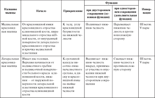
The end of the table. 10
Typical features of the masticatory muscles
The superficial layer of the masseter muscle in brachycephaly and hameprosopic face is usually wide and low, muscle fibers diverge downward (Fig. 85); with dolichocephaly and leptoprosopic face shape, it is long and narrow, muscle fibers run parallel. The intermediate layer of this muscle with dolichocephaly and leptoprosopia protrudes more from under the posterior edge of the superficial layer than with brachycephaly and hameprosopia.
The temporalis muscle with the dolichocephalic shape of the skull is low and long, and with the brachycephalic one - high and short (see Fig. 85).
Both heads of the lateral pterygoid muscle with the brachycephalic shape of the skull are short and wide, with a narrow gap between them, with the dolichocephalic muscle - long and narrow, with a wide gap between them (Fig. 86).
The medial pterygoid muscle with the dolichocephalic shape of the skull and the leptoprosopic shape of the face is long and narrow, and with brachycephaly and chameprosopia it is low and wide (Fig. 87).
The shape of the pterygoid and masticatory muscles is determined by the shape of the mandible branch and infratemporal fossa, but at the same time it corresponds to the structure of the bone components of the temporomandibular joint. This connection is especially clearly reflected in the external structure of the lateral pterygoid muscle. When the mouth is opened (lowering the lower jaw) and when the lower jaw is extended forward in people with a brachycephalic skull, the head of the joint is displaced to the apex of the flat articular tubercle, i.e. the articular path deviates slightly from the horizontal plane. This movement of the jaw head is provided by the lower head of the lateral pterygoid muscle, which lies almost horizontally. With the dolichocephalic shape of the skull, the articular head slides down the steep and high slope of the articular tubercle rather than horizontally. This movement is provided by the lower head of the lateral pterygoid muscle, the origin of which is located lower on the high lateral plate of the pterygoid process, and the muscle pulls the jaw head down rather than forward.